Monday, March 23, 2009

Nuclear E-cadherin and VHL Immunoreactivity are Prognostic Indicators of Clear-cell Renal Cell Carcinoma.

Abstract and Introduction

Abstract

The loss of functional von Hippel-Lindau (VHL) tumor suppressor gene is associated with the development of clear-cell renal cell carcinoma (CC-RCC). Recently, VHL was shown to promote the transcription of E-cadherin, an adhesion molecule whose expression is inversely correlated with the aggressive phenotype of numerous epithelial cancers. Here, we performed immunohistochemistry on CC-RCC tissue microarrays to determine the prognostic value of E-cadherin and VHL with respect to Fuhrman grade and clinical prognosis. Low Fuhrman grade and good prognosis associated with positive VHL and E-cadherin immunoreactivity, whereas poor prognosis and high-grade tumors associated with a lack of E-cadherin and lower frequency of VHL staining. A significant portion of CC-RCC with positive VHL immunostaining correlated with nuclear localization of C-terminally cleaved E-cadherin. DNA sequencing revealed in a majority of nuclear E-cadherin-positive CC-RCC, subtle point mutations, deletions and insertions in VHL. Furthermore, nuclear E-cadherin was not observed in chromophobe or papillary RCC, as well as matched normal kidney tissue. In addition, nuclear E-cadherin localization was recapitulated in CC-RCC xenografts devoid of functional VHL or reconstituted with synthetic mutant VHL grown in SCID mice. These findings provide the first evidence of aberrant nuclear localization of E-cadherin in CC-RCC harboring VHL mutations, and suggest potential prognostic value of VHL and E-cadherin in CC-RCC.

Introduction

Renal cell carcinoma (RCC) accounts for approximately 3% of all adult malignancies, with the clear-cell type (CC-RCC) comprising 80% of RCC.[1,2] Approximately 25-30% of CC-RCC patients have metastatic disease at diagnosis, and 20-30% of patients with clinically localized CC-RCC develop metastasis post-nephrectomy.[3] Recent advances in the use of antiangiogenesis-targeted agents appear to have revolutionized treatment.[4,5,6,7] Currently, a prediction of patient survival is based on traditional clinical parameters, including tumor size and Fuhrman nuclear grade.[8] However, the emerging understanding of the molecular pathways implicated in CC-RCC genesis and progression is providing previously unappreciated markers, which may serve as additional or better prognostic indicators of CC-RCC.

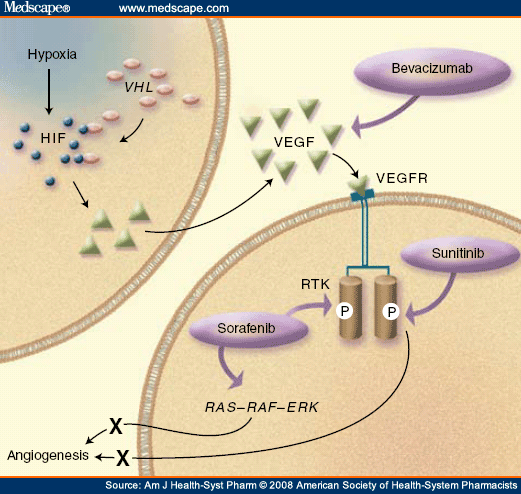
The principal cause of sporadic CC-RCC and familial von Hippel-Lindau (VHL) disease-associated CC-RCC is the inactivating mutations of VHL. Although VHL patients also develop tumors in other organs including the central nervous system, retina and the adrenal gland, CC-RCC remains to be the leading cause of morbidity and death for VHL patients.[9] The most well-established function of VHL is its role in the oxygen-dependent negative regulation of hypoxia-inducible factor (HIF). VHL is a substrate-conferring component of an E3 ubiquitin ligase ECV (elongins/Cul2/VHL) that polyubiquitylates the catalytic α-subunit of HIF that has undergone hydroxylation on conserved prolyl residues within the oxygen-dependent degradation (ODD) domain. Prolyl hydroxylation is mediated by a class of prolyl hydroxylases (PHD1-3) in an oxygen-dependent manner. Thus, under hypoxia or in the absence of a functional VHL, HIFα becomes stabilized and binds to its common and constitutively expressed β-subunit, forming an active HIF transcription factor capable of transactivating numerous hypoxia-inducible genes such as vascular endothelial growth factor (VEGF), glucose transporter-1 (GLUT1) and erythropoietin (EPO).[9] These discoveries have confirmed VHL as a critical regulator of the ubiquitous mammalian oxygen-sensing pathway.

HIFα is overexpressed in most tumors including CC-RCC. The re-introduction of VHL into VHL-null CC-RCC abrogates the tumorigenic potential of these cells in a mouse xenograft model.[10,11] Kondo et al[12] have shown that the expression of a non-degradable form of HIF2α was able to restore the tumorigenic phenotype in CC-RCC cells ectopically expressing VHL. Conversely, shRNA-mediated knockdown of HIF2α was sufficient to suppress the tumorigenic capacity of VHL-null CC-RCC cells.[13] Notably, the emerging evidence suggests that VHL mutations affecting HIF regulation were invariably associated with a subtype of VHL disease with a greater propensity of CC-RCC. Although these reports support the notion that deregulated accumulation of HIFα upon the loss of VHL is crucial for the development of CC-RCC, the precise downstream target(s) of HIF that are responsible for renal epithelial oncogenesis have, until recently, remained unclear.

The regulation of adhesive interactions between cells is critical during cell growth and differentiation, and the loss of cell-cell adhesion is frequently associated with tumor progression and metastasis.[14] Recently, VHL was shown to promote the transcription of E-cadherin via HIF-dependent activation of E-cadherin-specific transcriptional repressors.[15,16,17] Thus, a loss of VHL in CC-RCC results in the hyperactivation of HIF that triggers the expression of E-cadherin repressors, which in turn attenuates the expression of E-cadherin.[15,16,17] E-cadherin, a homophilic adhesion molecule associated with catenins that functions as a major component of cell junctions in polarized epithelial cells, is an established tumor suppressor. The graded loss of E-cadherin correlates with the aggressiveness of numerous carcinomas and the worsening of prognosis, while forced expression of E-cadherin suppresses tumor development and invasion in various in vitro and in vivo tumor model systems.[14]

The full-length E-cadherin (120 kDa) can be proteolytically cleaved at a cleavage site near the transmembrane domain, which releases an extracellular N-terminal 80-kDa fragment and generates a 38-kDa C-terminal fragment that can be further processed into a 33-kDa soluble cytosolic fragment.[18] The proteolytic ectodomain release or 'shedding' of E-cadherin is emerging as an important regulatory mechanism and has been suggested to cause rapid changes in cell adhesion, signaling and apoptosis.[19,20,21,22] Furthermore, increased levels of the soluble N-terminal fragment have been associated with several tumors, including prostate,[23] gastric,[24] hepatocellular[24] and bladder cancer and may be of prognostic value.[21] Whether the C-terminal intracellular fragment of E-cadherin has oncogenic roles, for example, via promoting β-catenin-mediated signaling, is unclear.

Here, we examined the prognostic value of VHL and E-cadherin in CC-RCC. While CC-RCC with negative E-cadherin staining exhibited no VHL immunoreactivity as expected, a significant portion of CC-RCC had aberrant nuclear E-cadherin staining despite positive VHL staining. DNA sequencing revealed in a majority of the nuclear E-cadherin-positive CC-RCC, subtle point mutations, deletions and insertions in VHL. The nuclear E-cadherin was not observed in chromophobe or papillary RCC, suggesting this to be specific to CC-RCC. Low Fuhrman grade and better prognosis associated with positive VHL and nuclear E-cadherin immunoreactivity, whereas high-grade tumors associated with a lack of nuclear E-cadherin staining and lower frequency of VHL staining. These findings provide the first evidence of an aberrant nuclear localization of E-cadherin in CC-RCC harboring VHL mutations, and suggest potential prognostic value of VHL and E-cadherin status in CC-RCC.

Materials and Methods

Cell Culture

Human 786-O CC-RCC cells were cultured in Dulbecco's modified Eagle's medium containing 10% heat-inactivated fetal bovine serum (Sigma, Saint Louis, MI, USA) at 37°C in a humidified 5% CO2 atmosphere. 786-O subclones stably expressing hemagglutinin (HA)-tagged wild-type VHL30, HA-VHL19(WT), HA-VHL19(MUT) with V84M and P192Q mutations, or empty plasmid were generated as previously described.[25,26]

Antibodies

Anti-HA (12CA5; Roche Applied Science, Basel, Switzerland), anti-TCP1 (Abcam Inc., Cambridge, MA, USA), C-terminal-specific anti-E-cadherin (BD Biosciences, Mississauga, ON, Canada), N-terminal-specific anti-E-cadherin (Vector laboratories, Burlingame, CA, USA), anti-VHL (IG32) (BD Biosciences, Mississauga, ON, Canada), and anti-β-catenin (BD Biosciences, Mississauga, ON, Canada) monoclonal antibodies and anti-Glut1 polyclonal antibodies (Alpha Diagnostic International Inc., San Antonio, TX, USA) were obtained from the companies indicated.

Plasmids

The mammalian expression plasmid pRc-CMV-HA-VHL30 was previously described.[25,26] pRc-CMV-HA-VHL19(WT) was generated from the pRc-CMV-HA-VHL30 plasmid using primer set 5'-CCCAAGCTTATGTATCCATATGATGTTCCAGATTATGCTGAGGCCGGGCGGCCG-3'/5'-GCTCTAGATCAATCTCCCATCCGTTGATGTGC-3'. pRc-CMV-VHL19(MUT), V84M and P192Q, was serendipitously generated from pRc-CMV-HA-VHL30 using the above primers in a PCR reaction. The authenticity of the plasmids was confirmed by direct DNA sequencing.

Immunoprecipitation and Immunoblotting

Cells were lysed in EBC buffer (50 mM Tris (pH 8), 120 mM NaCl, 0.5% NP-40) supplemented with protease and phosphatase inhibitors (Roche, Laval, QC, Canada). Samples were supplemented with the indicated antibody and immobilized on protein A-Sepharose beads (Amersham Biosciences, Uppsala, Sweden). The samples were then washed five times with NETN buffer (20 mM Tris (pH 8), 120 mM NaCl, 1 mM EDTA, 0.5% NP-40), eluted by boiling in sodium dodecyl sulfate (SDS)-containing sample buffer, separated by SDS-polyacrylamide gel electrophoresis (PAGE) and electrotransferred to a polyvinylidene difluoride membrane (Bio-Rad, Mississauga, ON, Canada). Specific protein bands on Western blots were visualized using the various antibodies indicated. All primary antibodies were diluted in phosphate-buffered saline (PBS) containing 0.1% Tween 20. Horseradish peroxidase-conjugated goat anti-mouse, goat anti-rabbit or rabbit anti-goat antibody (Pierce, Rockford, IL, USA) was used at 1:20 000 dilution as a secondary antibody, after which enhanced chemiluminescence (PerkinElmer Life Sciences, Woodbridge, ON, Canada) was performed for detection.

Metabolic Labeling

Metabolic labeling was performed as previously described.[27] Briefly, radioisotope labeling was performed by methionine starvation for 45 min, followed by growth in 5 ml of methionine-free Dulbecco's modified Eagle's medium supplemented with [35S]methionine (100 μCi/ml of medium; PerkinElmer Life Sciences, Woodbridge, ON, Canada) and 2% dialyzed fetal bovine serum at 37°C in a humidified 5% CO2 atmosphere for 3 h for the interaction studies and 1 h for half-life analysis.

RNA Extraction

RNA was extracted from cells using RNeasy kit according to the manufacturer's instructions (Qiagen, Mississauga, ON, Canada).

Quantitative Real-time PCR

Quantitative real-time PCR was performed as previously described.[28] Briefly, first-strand cDNA synthesis was performed as follows: 1 μl of oligo(dT)23 primer (Sigma, St Louis, MI, USA) was incubated with 5 μg of RNA and dH2O (total reaction volume was 20 μl) for 10 min at 70°C in a thermal cycler (MJ research, Boston, MA, USA). The mixture was cooled to 4°C, at which time 4 μl of 5 × first-strand reaction buffer, 2 μl of 0.1 M DTT, 1 μl of 10 mM dNTPs and 1 μl Superscript II reverse transcriptase (Invitrogen, Carlsbad, CA, USA) were added. cDNA synthesis was performed for 1.5 h at 42°C, followed by 15 min at 70°C in the thermal cycler. Human genomic DNA (Roche, Mannheim, Germany) standards or cDNA equivalent to 20 ng of total RNA were added to the qPCR reaction in a final volume containing 1 × PCR buffer (without MgCl2), 3 mM MgCl2, 0.25 U Platinum Taq DNA polymerase, 0.2 mM dNTPs, 0.3 μl SYBR Green I, 0.2 μl ROX reference dye and 0.5 μM primers (Invitrogen, Carlsbad, CA, USA). Amplification conditions were as follows: 95°C (3 min); 40 cycles of 95°C (10 s), 65°C (15 s), 72°C (20 s), 60°C (15 s) and 95°C (15 s). qPCR was performed using the ABI Prism 7900HT Sequence Detection System (Applied Biosystems, Foster City, CA, USA). Gene-specific oligonucleotide primers designed using Primer Express (Applied Biosystems) were as follows: GLUT-1 primer set 1 (5'-CACCACCTCACTCCTGTTACTTACCT-3' and 5'-CAAGCATTTCAAAACCATGTTTCTA-3'), GLUT-1 primer set 2 (5'-CTCCCAGCAGCCCTAAGGAT-3' and 5'-ATCTGTCAGGTTTGGAAGTCTCATC-3') and β-actin primer set (5'-GGATCGGCGGCTCCAT-3' and 5'-CATACTCCTGCTTGCTGATCCA-3'). SYBR I fluoresces during each cycle of the qPCR by an amount proportional to the quantity of amplified cDNA (the amplicon) present at that time. The point at which the fluorescent signal is statistically significant above the background is defined as the cycle threshold (Ct). Expression levels of the various transcripts were determined by taking the average Ct value for each cDNA sample performed in triplicate, and measured against a standard plot of Ct values from amplification of serially diluted human genomic DNA standards. Since the Ct value is inversely proportional to the log of the initial copy number, the copy number of an experimental mRNA can be obtained from linear regression of the standard curve. A measure of the fold difference in copy number was determined for each mRNA. Values were normalized to the expression of β-actin mRNA and represent the average value of both GLUT-1 primers in three independent experiments performed in triplicate.

In Vitro Ubiquitination Assay

An in vitro ubiquitination assay was performed as previously described.[29] The [35S]methionine-labeled reticulocyte lysate HA-HIF-1α ODD translation product (4 μl) was incubated in 786-O RCC S100 extracts (100-150 μg) supplemented with 8 μg/μl ubiquitin (Sigma), 100 ng/μl ubiquitin aldehyde (Boston Biochem Inc., Cambridge, MA, USA) and an ATP-regenerating system (20 mM Tris (pH 7.4), 2 mM ATP, 5 mM MgCl2, 40 mM creatine phosphate and 0.5 μg/μl creatine kinase) in a reaction volume of 20-30 μl for 1.5 h at 30°C.

Protease Digestion Assay

Protease sensitivity was assayed as previously described.[30] Briefly, lysates were incubated with chymotrypsin (10 μg/ml) for 10 min on ice. Reactions were stopped by the addition of 1 mM PMSF before immunoprecipitation (IP) and SDS-PAGE.

SCID Mouse Xenograft Assay

Multiple 786-O subclones expressing HA-VHL30, HA-VHL19(MUT) or empty plasmid were grown to ˜ 90% confluence in a humidified 5% CO2 atmosphere at 37°C. Cells were harvested with a solution of 0.25% trypsin and 1 mM EDTA. Cells (1 × 10⁷) in 100 μl of 1 × PBS were injected intramuscularly into the left hind legs of male SCID mice (Charles River Laboratories Inc., Wilmington, MA, USA). Tumor growth was assessed and measured weekly by carefully passing the tumor-bearing leg through a series of holes of decreasing diameter (0.5-mm decrements) in a plastic rod.

Immunohistochemistry

SCID mouse xenograft tumors were fixed in 10% neutral buffered formalin and later embedded in paraffin. Formalin-fixed, paraffin-embedded sections from 19 nephrectomy specimens with CC-RCC were obtained from the Department of Pathology and Laboratory Medicine at The University Health Network (Toronto, ON, Canada). These tissue blocks were used and processed in accordance with a University Health Network Research Ethics Board-approved protocol concerning gene expression in RCC. Tissues were fixed in 10% neutral buffered formalin for 24-36 h. Representative sections of tumor with adjacent non-tumor renal parenchyma, 3-4 mm in thickness, were embedded in paraffin and 5-μm sections were cut and placed on coated slides for light microscopy.

Tumor morphology and classification were assessed using standard hematoxylin and eosin (H&E) staining. The tumors were classified as conventional clear-cell type according to criteria described in the WHO classification of renal tumors.[31] Immunohistochemical staining for VHL was performed manually using a standard avidin-biotin-peroxidase complex method. Briefly, unlabeled anti-HA, anti-Glut1, anti-E-cadherin (BD biosciences, Mississauga, ON, Canada), anti-E-cadherin (Vector laboratories, Burlingame, CA, USA) and anti-β-catenin were used at a 1:2000 dilution, and an overnight incubation following microwave pretreatment for antigen retrieval. The sections were then incubated with a biotinylated secondary antibody (horse anti-mouse IgG, 1:200 dilution) and the avidin-peroxidase complex. The colored reaction was visualized using nova red as the chromogen. The tissue was then mildly counterstained with hematoxylin.

Tissue Microarray

Tissue microarray (TMA) consisting of quadruplicate representative 1.0-mm cores from 56 CC-RCC with various Fuhrman grades (grades 1-4) and stages (pT1-pT4) were used to analyze the correlation between E-cadherin and VHL protein expression. Sections (5 μm) from the CC-RCC TMA were stained with anti-E-cadherin and anti-VHL antibodies as described above. The slides were then scanned using a ScanScope (Aperio Technologies, Vista, CA, USA) and scored by two observers. In order to be considered for evaluation, at least half of the TMA core was required to be present on the slide, with at least 50% of the tissue present being tumor cells. Cores were being scored as either positive or negative. Only tumors with the majority of cores in agreement (3 out of 4 or 2 out of 3) were used for further analysis.

VHL Sequencing

VHL was amplified from patient samples using primers 5'-CAGTAACGAGTTGGCCTAGC-3'/5'-GGATGTGTCCTGCCTCAAG-3' for exon 1, 5'-GGACGGTCTTGATCTCCTGA-3'/5'-GATTGGATACCGTGCCTGAC-3' for exon 2 and 5'-CTGCCACATACATGCACTCA-3'/5'-AAGGAAGGAACCAGTCCTGT-3' for exon 3. PCR reactions were performed using the Qiagen Multiplex Master Mix (Qiagen, Mississauga, ON, Canada) according to manufacturer's instructions. Exons were sequenced with the Cy5/Cy5.5 Dye Primer Cycle Sequencing kit (Amersham, Quebec/Visible Genetics, Toronto) according to the manufacturer's instructions. Each primer mixture contained two primers, labeled with either Cy5 or Cy5.5. Primers used for sequencing were 5'-/5Cy5/-CTAGCCTCGCCTCCGTTA-3'/5'-/5Cy5/-GCCTCAGTTCCCCGTCTG-3' for exon 1, 5'-/5Cy5-GATACCGTGCCTGACATCAG-3' for exon 2 and 5'-/5Cy5/-CAGGTAGTTGTTGGCAAAGC-3' for exon 3. Two primers were used for exon 1 as it is a large exon. The sequences generated were compared with wild-type VHL (GenBank accession number AC_000046, for sequence alterations, using the OpenGene Automated DNA System and Gene Librarian software, version 3.1 (Visible Genetics, Suwanee, GA, USA).

Statistics

Fisher's exact test was used to determine whether positive VHL immunoreactivity and nuclear E-cadherin subcellular localization were interdependent. Two-tailed P-values less than 0.05 were considered significant.

Role of Sunitinib and Sorafenib in the Treatment of Metastatic Renal Cell Carcinoma

Abstract and Introduction
Abstract

Purpose: The role of sunitinib and sorafenib in the treatment of metastatic renal cell carcinoma (mRCC) is reviewed.
Summary: Sunitinib malate is a potent inhibitor of vascular endothelial growth factor (VEGF) receptors, FMS-like tyrosine kinase 3 (FLT3), c-KIT, and platelet-derived growth factor (PDGF), which give the drug its direct antitumor and antiangiogenic properties. Sunitinib is currently approved as a second-line treatment of mRCC in patients who have either not responded to or who are not eligible to receive interleukin-2. Clinical trials of sunitinib have found similar rates of partial response, disease stabilization, and progression-free survival. Sorafenib inhibits VEGF receptors, PDGF receptors, FLT3, RAF-1, and BRAF in vitro and has been shown to prevent the growth of tumors but not to reduce tumor size. Sorafenib has been proven to improve survival in a novel randomized discontinuation trial and a Phase III randomized, placebo-controlled trial. No studies have directly compared the effectiveness of sunitinib to sorafenib in the treatment of advanced renal cell carcinoma. Sunitinib and sorafenib share a similar mechanism of action and primarily target tumor angiogenesis by inhibiting a variety of tyrosine kinases; the agents have similar toxicity, with the exception of an increased risk of hypertension associated with the use of sorafenib. Sorafenib does not result in tumor shrinkage, but sunitinib significantly reduces tumor size.
Conclusion: The tyrosine kinase inhibitors sorafenib and sunitinib offer improved outcomes for patients with mRCC, but they are far short of a cure. Despite the introduction of sorafenib and sunitinib, palliative care is still an acceptable treatment option for mRCC because of the disease's extremely poor prognosis.
Introduction

In 2007, 51,190 patients in the United States were expected to be diagnosed with kidney cancer, accounting for 2% of all cancers.[1] Renal cell carcinoma (RCC) comprises 90% of all kidney cancers. Patients at a high risk for developing RCC include heavy smokers, urban dwellers, and patients with genetic predispositions, such as Von Hippel-Lindau (VHL) disease, type 2 papillary RCC, and Birt-Hogg-Dube syndrome.[2] Other possible risk factors include polycystic kidney disease, diabetes mellitus, and chronic dialysis. The median age at RCC diagnosis is 65 years.[2] In two thirds of patients diagnosed with nonmetastatic RCC, the tumor is found incidentally. Only 10% of patients complain of RCC's main symptoms—flank pain, flank mass, and hematuria—until advanced stages of the disease.[2] Since symptoms are uncommon with early disease, many patients who seek medical advice have already developed metastatic RCC (mRCC). Common sites of metastases in these patients include the lungs, liver, bones, and brain.

Prognosis is based on stage as determined by the Tumor Node Metastasis staging classification. Patients without metastasis—stage I, II, or III RCC—have a predicted five-year survival rate of 91%, 74%, or 67%, respectively.[3] Even if the tumor has extended into the renal vein, the five-year survival rate is still 25-50%.[2] Early-stage RCC is potentially curable with nephrectomy.

Despite the effectiveness of a nephrectomy in treating RCC, 20-30% of patients' RCC will progress to mRCC.[4] Progression to metastatic disease within one year after nephrectomy confers the same survival rate as stage IV disease without nephrectomy.[2] In patients with stage IV disease, the median survival time is 10 months, and the one-, two-, and three-year survival rate is 42%, 20%, and 11%, respectively.[5] If metastatic disease develops more than two years after nephrectomy, then the patient has a more favorable prognosis with a five-year survival rate of 20%.[2] Nephrectomy is generally discouraged in patients with mRCC, as less than 1% of patients who undergo nephrectomy experience spontaneous remission and any benefit is far exceeded by the risk of surgery.[2]

RCC histologies are characterized by the cell of origin, morphology, and growth pattern. Five RCC subtypes have been identified: clear-cell, chromophilic, chromophobic, oncocytic, and collecting-duct tumors. The most common RCC histology is clear cell, accounting for 85% of all RCCs.[6] Clear-cell RCC is associated with a mutation in the gene that encodes for the VHL protein. VHL is a tumor-suppressor protein whose gene product acts like an antiangiogenic agent, inhibiting the actions of various hypoxia-induced genes. Individuals with VHL disease have a somatic or inherited mutation and do not produce the VHL protein. Inherited VHL disease occurs in every 1 of 36,000 births, and these individuals have a 30% chance of developing RCC, along with other systemic problems.[7] More commonly, a somatic mutation occurs, causing the VHL protein to be ineffective. Left unchecked, the hypoxia-inducible factors 1α and 2α promote the release of vascular endothelial growth factor (VEGF) and platelet-derived growth factor (PDGF), even in the presence of normoxia (Figure 1).[8] This allows angiogenesis and new vessel growth to proceed in the absence of hypoxia and injury.
http://images.medscape.com/images/571/655/art-ajh571655.fig1.gif

Due to the inactivation of VHL, overexpression of VEGF and PDGF, and increase in angiogenesis, clear-cell RCC is characterized by highly vascular primary tumors and metastases. Currently available treatment options, including chemotherapy, interleukin-2 (IL-2), interferon-alfa (IFN-α), and a combination of IL-2 and IFN-α, typically result in response rates of 5%, 20%, 12%, and 19%, respectively ( Table 1 ).[18-21] IL-2 is currently approved for the treatment of advanced RCC. Fourteen percent of patients achieve a complete response, the median duration of which has not been reached.[19] IL-2 is a first-line treatment for advanced RCC;[22] however, IL-2 cannot be given to many patients with advanced RCC because of significant toxicities that require drug administration and monitoring in an intensive care setting.[19] The National Comprehensive Cancer Network (NCCN) recommends palliative care as a first-line treatment option for patients with mRCC because of the limited and ineffective treatment options available for mRCC.[22]

Recently, angiogenesis inhibitors have been studied in the treatment of clear-cell mRCC. Bevacizumab, a monoclonal antibody that specifically targets soluble VEGF, has demonstrated improved progression-free survival time versus placebo but failed to show a benefit in overall survival in a Phase II randomized trial.[23] Bevacizumab is currently a second-line treatment for clear-cell mRCC.[22] Because of bevacizumab's promising results, other agents that target both VEGF and PDGF have been studied for the treatment of advanced RCC.

Unlike bevacizumab, which targets extracellular VEGF, tyrosine kinase inhibitors target the intracellular signaling pathways of VEGF receptors. Figure 1 shows how VEGF increases angiogenesis and the sites of action of various antiangiogenic agents. Tyrosine kinase inhibitors target VEGF receptors and a variety of receptors that rely on a tyrosine kinase component to function properly, including PDGF, FMS-like tyrosine kinase 3 (FLT3), RAF, and c-KIT.[24] The receptor structure consists of an extracellular binding site, a transmembrane region, and an intracellular tyrosine kinase domain. Binding-site activation induces stabilized receptor dimerization, which leads to increased tyrosine kinase activity. This catalyzes a series of reactions and signaling pathways that generally lead to proliferation.[10] The small-molecule tyrosine kinase inhibitors sorafenib and sunitinib inhibit the receptor activation.

A University of Chicago Consortium Phase II Trial of SB-715992 in Advanced Renal Cell Cancer

Abstract and Introduction
Abstract

Background: Advanced renal cell cancer (RCC) continues to have a poor overall prognosis despite new FDA-approved therapies. Although taxane-based therapies are generally ineffective in RCC, research into the role of the von Hippel-Lindau protein has shown an association with microtubule dynamics. Mitotic kinesins are a class of molecular motors that also interact with microtubules and are required for proper mitotic function. SB-715992 is a new agent that inhibits the function of a mitotic kinesin known as kinesin spindle protein and leads to cell death.
Patients and Methods: Twenty patients with previously treated advanced RCC were enrolled on this phase II trial of SB-715992, with response rate as a primary endpoint.
Results: No patients responded with complete or partial remission. Six patients had stable disease, and 1 patient continues on therapy after 12 cycles. Common toxicities included anemia (80%), elevated creatinine (70%), lymphopenia (45%), fatigue (50%), hyperglycemia (50%), and dyspnea (45%). Reported grade 3/4 toxicities included dyspnea, fatigue, neutropenia with skin infection, dizziness, hyperuricemia, and hypertension.
Conclusion: This dose and schedule of SB-715992 does not appear to have a significant cytotoxic effect for patients with previously treated advanced RCC.
Introduction

The American Cancer Society anticipated that over 51,000 new cases of renal cell carcinoma (RCC) would be diagnosed in the United States in 2007, and almost 13,000 patients would die of this disease.[1] Incidence and mortality have steadily increased over time.[2] An estimated 20% of patients will have locally advanced disease at diagnosis, and up to 40% of patients treated by nephrectomy for localized disease will relapse.[3] Another 25% will have metastatic disease at diagnosis. The prognosis for recurrent or metastatic disease is poor, with a 5-year survival rate of < 10%, but individual patient outcome is highly variable, with median survival of 20 months in good-prognosis, 10 months in intermediate-prognosis, and 4 months in poor-prognosis patients.[4] In addition, there is increasing recognition that RCC is composed of multiple histologic subtypes with distinct pathologic and biologic characteristics, of which clear cell is the most common. Although high-dose interleukin-2 offers long-term survival to a small percentage of patients with clear-cell RCC, the majority of patients are not candidates for this relatively toxic approach.[5] More recently, antiangiogenic therapies have been shown to significantly increase progression-free survival in patients with good- and intermediate-prognosis clear-cell disease.[6-9] The mammalian target of rapamycin inhibitor temsirolimus has also been shown to improve survival of patients with poor-prognosis RCC.[10] These therapies, however, are not curative. Thus, alternative treatments are still needed.

Targeting the mitotic spindle is one such approach. Recent evidence has demonstrated that the von Hippel-Lindau protein (pVHL), which is mutated or methylated in the majority of clear-cell RCC, is associated with microtubule function.[11,12] The taxanes are classic spindle targeting agents that bind to microtubules and modify microtubule polymer dynamics. These agents are also known to be ineffective in the treatment of RCC. The mechanism of resistance to taxanes in RCC has not yet been fully elucidated. However, it might be related to alterations in the expression of tubulin isotypes or enhanced expression of the multidrug resistance-related transporter efflux pumps such as P-glycoprotein and multidrug resistance associated protein 2.[13,14] The epothilone ixabepilone, which also binds to microtubules, has shown some promising activity in RCC.[15,16] Another mitotic spindle protein is the mitotic kinesin spindle protein (KSP). This protein plays an exclusive and essential role in assembly and function of the mitotic spindle. Kinesin spindle protein expression is higher in many cancer tissues compared with adjacent normal tissue and thus represents a novel target for cancer treatment. Additional data suggests that KSP inhibitors might be effective in taxane-resistant cells.[17,18]

SB-715992 is a polycyclic, nitrogen-containing heterocyclic inhibitor of KSP, and it is the first of its class to enter clinical trials. This agent blocks assembly of the functional mitotic spindle, thereby causing cell-cycle arrest in mitosis and subsequent cell death. Preclinical models have shown a broad spectrum of activity against cancer, including models that are refractory to cytotoxic chemotherapy. Several phase I studies of SB-715992 have already been conducted, and the dose-limiting toxicity of the weekly (7 mg/m2) and every-21-day regimens (18 mg/m2) is neutropenia.[19-21] Other toxicities include constipation, fatigue, and transaminitis. Given the association of pVHL with microtubule function and the overall safety profile to date, including the absence of neuropathy, further study of this agent for RCC is warranted.Patients and Methods
Patient Eligibility Criteria

Patients aged < 18 years were eligible if they met the following conditions: Eastern Cooperative Oncology Group performance status ≤ 2, histologically or cytologically confirmed metastatic RCC or unresectable primary tumor, a minimum of 1 but no more than 2 previous therapies in the 8 months before enrollment, < 28 days since previous treatment, absolute granulocyte count ≥ 1500 cells/mm3, hemoglobin ≤ 9 mg/dL, platelet count ≥ 100,000 cells/mm3, total bilirubin < 2 mg/dL, aspartate aminotransferase and alanine aminotransferase ≤ 2.5 × institutional upper limit of normal, serum creatinine ≤ 2.0 or calculated creatinine clearance ≥ 40 mL per minute, and corrected QT interval of < 0.47 seconds. Patients were excluded for any of the following reasons: if they had received previous tubule, DNA, or mitosis targeting agents for the treatment of RCC; if they were pregnant or nursing women; if they were HIV positive; or if they had a history of brain metastases. Because SB-715992 is an in vitro inhibitor of CYP3A4, medications or substances that are known as significant inhibitors or inducers of CYP3A4 were prohibited within 14 days (< 6 months for amiodarone) before the administration of the first dose of SB-715992. All patients were required to provide written informed consent according to federal, state, and institutional guidelines.
Treatment Plan

SB-715992 was administered at 7 mg/m2 intravenously on days 1, 8, and 15, every 28 days. Patients who experienced any response or stable disease (SD) continued protocol treatment until progression, unacceptable toxicity, intercurrent illness, or delay of treatment for < 3 weeks for any reason. For grade 4 neutropenia or thrombocytopenia lasting < 4 days, grade 3/4 neutropenia associated with fever, nonhematologic toxicity of grade ≤ 3, or grade 2 neurotoxicity, dose reductions were made by 1 mg/m2 increments up to a minimum dose of 5 mg/m2. Grade 3 or greater neurotoxicity resulted in the removal of the patient from protocol treatment.
Patient Evaluation

Patients were required to have a clinical visit and laboratory tests done within 7 days of registration. In addition, all baseline radiographic studies were completed within 4 weeks of registration. Disease status was assessed according to RECIST (Response Evaluation Criteria for Solid Tumors) every 8 weeks.[22]
Statistical Analysis

The primary objective of this phase II trial was to evaluate the objective response rate to SB-715992 in patients with metastatic RCC. An optimal 2-stage accrual design was implemented with a null hypothesis that SB-715992 would have a ≤ 10% true response rate.23 The alternative hypothesis would be a true response of ≥ 30%, and a and ß errors of 0.05 and 0.1, respectively, was adopted. Initially, 18 patients were to be accrued, with expansion to a total of 35 if > 2 patients responded. Further evaluation of this agent would then be recommended if ≥ 7 of the 35 eligible patients demonstrated a response. Secondary analysis included evaluation of toxicity, including overall and type of toxicity.

Immunohistochemical Detection of the von Hippel-Lindau Gene Product (pVHL) in Human Tissues and Tumors: A Useful Marker for Metastatic Renal Cell Carc

Abstract and Introduction
Abstract

Genetic alteration of the von Hippel-Lindau (VHL) tumor suppressor gene has been linked to hereditary and sporadic clear cell renal cell carcinomas (RCCs). Inconsistent data on immunodetection of the VHL gene product (pVHL) in normal tissues and tumors have been reported. We immunohistochemically reevaluated the usefulness of a specific rabbit polyclonal anti-pVHL antibody in 531 cases of renal and nonrenal neoplasms and normal tissues. Positive immunostaining was observed in nearly 100% of primary renal neoplasms, 95% of metastatic RCCs, and 90% of clear cell carcinomas of the ovary and uterus. In normal tissues, positive immunoreactivity was observed only in renal tubules, exocrine pancreas, islets, and bile ducts. Western blot and reverse transcription–polymerase chain reaction confirmed the immunostaining results. These data indicate that this anti-pVHL antibody is a useful marker in assisting diagnosis of metastatic RCC and may serve as a diagnostic marker for clear cell carcinomas of the ovary and uterus.
Introduction

Renal cell carcinoma (RCC) is the ninth most common malignant tumor in the United States, accounting for approximately 2% of all cancers. In 2006, more than 38,890 new cases of RCC were diagnosed, and an estimated 12,840 people died of this cancer in the United States alone.[1,2] When patients have local disease at initial examination, a favorable prognosis is usually anticipated; however, metastatic RCC has a much worse prognosis.[2] Identification of a metastatic RCC is not always straightforward because RCC is notorious for distant metastases years after the initial diagnosis and treatment. In addition, the morphologic complexity of RCC may create a diagnostic challenge. According to the World Health Organization classification,[3] RCC is subdivided into clear cell, chromophobe, papillary, collecting duct, multilocular cystic renal cell, medullary, mucinous tubular spindle cell, and unclassified RCC. Because of their histologic variants and morphologic complexity, RCCs frequently show histologic features that resemble many other neoplasms. It can be challenging to distinguish them from other metastatic malignancies based on morphologic features alone, especially when a specimen is limited, as in small tissue core biopsies or fine-needle aspiration biopsy specimens.

Many attempts have been made to identify sensitive and specific markers for RCCs. Some successes have been achieved. Coexpression of epithelial membrane antigen and vimentin is frequently present in clear cell RCC.[4] CD10 was detected in approximately 90% of RCCs, but also in many hepatocellular carcinomas, prostatic carcinomas, and urothelial carcinomas.[5,6] The RCC has a diagnostic sensitivity of 84% to 92% in primary RCC and 67% to 84% in metastatic RCC.[5,7,8] Carbonic anhydrase IX (MN/CA9), a tumor-associated antigen, was positive in 100% of clear cell RCCs and papillary RCCs, but it was also expressed in cervical dysplasia and carcinoma.[9] P504S, initially considered to be a specific marker for prostatic adenocarcinoma, has been shown to be positive in 100% of papillary RCCs and some primary and metastatic clear cell RCCs.[10-14] In addition, glutathione S-transferase α and carbonic anhydrase II were demonstrated to be specific markers for clear cell RCC and chromophobe RCC, respectively.[15] Expression of S-100 protein has been shown in 70% of clear cell RCCs but also was positive in many nonrenal tumors.[16]

Recently, we introduced human kidney injury molecule-1 (KIM-1), also known as TIM-1 (T-cell immunoglobulin domain and mucin domain protein 1) or HAVCR1 (hepatitis A virus cellular receptor), for diagnosing RCCs.[17] Our data showed that expression of KIM-1 was present in approximately 75% of primary and metastatic clear cell RCCs and nearly 100% of papillary RCCs. Most clear cell carcinomas of the ovary were also positive for KIM-1.[17] In contrast with previously published findings, KIM-1 is probably one of the most promising biomarkers, with the highest specificity in diagnosing RCCs. With a diagnostic sensitivity of 75%, it is useful but not ideal for detecting metastatic RCCs; therefore, we continue searching for a more sensitive marker.

Inactivation of the von Hippel-Lindau (VHL) tumor suppressor gene on chromosome 3p25-26 by mutation, deletion, or hypermethylation is a frequent event in hereditary and sporadic clear cell RCC.[2,18,19] Genetic alteration of the VHL gene seems to be a crucial step in the initiation and progression of clear cell RCC.[2,20] RCC cell lines with biallelic VHL inactivation form RCC when injected into the skin of nude mice; however, the tumors abrogate when VHL function is restored, which indicates that loss of VHL has a role in the initiation and maintenance of the tumor.[21]

The VHL gene encodes 2 biologically active proteins (pVHL) with molecular weights of 30 kd (pVHL30) and 19 kd (pVHL19).[22] The short form of pVHL seems to arise as a result of alternative translation initiation. Only limited data are available on the immunolocalization of pVHL in RCCs, normal tissues, and nonrenal tumors.[23-28] Notably, some of the data are inconsistent with regard to the compartmentalization and distribution of pVHL. Some technical challenges are also encountered.[23] Corless and coworkers[23] reported cytoplasmic staining of VHL in normal epithelial cells of many organs and various types of carcinomas, including clear cell RCC and carcinoma of the lung, prostate, colon, breast, bladder, and thyroid on frozen sections; however, the immunostains did not work on formalin-fixed, paraffin-embedded sections. Schraml et al[27] showed cytoplasmic localization of the VHL protein when using antibody to pVHL30, but nuclear and cytoplasmic staining for the VHL protein was noted when using antibody to pVHL30 and pVHL19.

In the present study, we used a well-characterized rabbit polyclonal antibody against the full-length VHL protein of human origin, attempting to reevaluate the expression pattern, the distribution in normal tissues, and the diagnostic usefulness of pVHL in a large series of specific subtypes of renal cell tumors, metastatic RCCs, and nonrenal tumors, using formalin-fixed, paraffin-embedded tissue microarray (TMA) and routine tissue sections along with Western blots and reverse transcription–polymerase chain reaction (RT-PCR). Our data not only confirmed and expanded the findings in some of the previous reports but also, more important, revealed the additional usefulness of this particular rabbit polyclonal anti-pVHL antibody in assisting diagnosis of metastatic RCC and detecting clear cell carcinoma of the ovary and uterus.Materials and Methods
Case Selection

The study was approved by the institutional review boards at Geisinger Medical Center, Danville, PA, and the coauthors' institutions. We retrieved 526 cases of renal epithelial neoplasms and nonrenal tumors from the archives of the Department of Laboratory Medicine, Geisinger Medical Center, and the departments of pathology from coauthors' institutions. Tissue samples were made anonymous and disassociated from any clinical data. TMA sections and routine large tissue sections were used in the study. The TMA sections included 80 conventional (clear cell) RCCs, 57 papillary RCCs, 15 chromophobe RCCs, 14 renal oncocytomas, 24 cases of nonneoplastic renal cortical-medullary tissue, and 94 nonrenal carcinomas. Cases in TMA sections were different from cases in routine sections. Routine sections from 37 metastatic clear cell RCCs, 11 chromophobe RCCs, 10 oncocytomas, and 213 nonrenal carcinomas from various organs were also included. Nonneoplastic tissues adjacent to the tumors were evaluated as well.
TMA Construction

The H&E-stained sections for the selected cases were reviewed, and the targeted areas were marked on the H&E-stained sections and the corresponding paraffin blocks. One 1.5-mm tissue core for each case was removed using a manual microarray device (Beecher Instruments, Sun Prairie, WI) and inserted into recipient paraffin blocks as described.[15,29]
Immunohistochemical Analysis Using a Rabbit Polyclonal Anti-pVHL Antibody

Immunohistochemical stains were performed on formalin-fixed, paraffin-embedded 4-µm sections of the TMA and routine tissue sections. The tissue sections were deparaffinized, and antigen retrieval was carried out in Proteinase K solution for 10 minutes at room temperature. Six antigen retrieval methods were tested before the Proteinase K retrieval method was chosen (see the "Discussion" section). The sections were incubated in 3% hydrogen peroxidase for 5 minutes to quench endogenous tissue peroxidase. The tissue sections were then incubated with a rabbit polyclonal antibody against pVHL (VHL [FL181]:sc-5575, Santa Cruz Biotechnology, Santa Cruz, CA)[6,21] at a 1:50 dilution for 30 minutes at room temperature. The slides were stained in a DAKO automated immunostainer using a standard EnVision-HRP kit (DAKO, Carpinteria, CA) as previously described.[16] The EnVision-HRP detection kit is designed to significantly reduce or eliminate nonspecific endogenous biotin activity. Immunohistochemical reactions were developed with diamino-benzidine as the chromogenic peroxidase substrate, and slides were counterstained with hematoxylin. Normal kidney tissue served as the positive control sample; negative control samples included replacement of the primary antibody with nonimmune rabbit serum.

Two surgical pathologists (F.L. and H.L.) independently evaluated the immunostained TMA and regular tissue sections. The staining results were recorded in a semiquantitative manner as follows: negative, less than 5% of tumor cells stained; 1+, 6% to 25%; 2+, 26% to 50%; 3+, 51% to 75%; and 4+, more than 75%. The staining intensity was also assessed and recorded as follows: weak, light brown, fine, granular cytoplasmic/membranous staining; or strong, dark brown, granular cytoplasmic/membranous staining. Nuclear staining, if present, was also recorded as a positive result (only when >5% of cells stained).
Immunohistochemical Analysis Using a Mouse Monoclonal Anti-VHL Antibody

To further validate the immunohistochemical staining results from this rabbit polyclonal anti-pVHL antibody, we also performed immunostains on selected TMA sections of renal and nonrenal tumors and VHL-related tumors (hemangioblastoma and pheochromocytoma) using another commercially available mouse monoclonal anti-pVHL antibody. This monoclonal antibody was used in the study by Schraml et al.[27]

Immunohistochemical stains were performed on formalin-fixed, paraffin-embedded 4-µm histologic sections. Six antigen retrieval methods were carried out: (1) target retrieval solution buffer (water bath heating for 20 minutes; cooling for 20 minutes); (2) TUF buffer (water bath heating for 10 minutes; cooling for 10 minutes); (3) EDTA (microwave heating for 20 minutes; cooling for 20 minutes); (4) citrate buffer (microwave heating for 20 minutes; cooling for 20 minutes); (5) Proteinase K, 10 minutes; and (6) no special treatment.

The staining procedure was the same as for the polyclonal antibody. The tissue sections were then incubated with the mouse monoclonal antibody to pVHL (catalog No MS-690-P, clone Ig33, dilution 1:50; LabVision, NeoMarkers, Fremont, CA). The slides were stained in a DAKO automated immunostainer using a standard EnVision-HRP detection kit as previously described.
Western Blot

Two Western blots were performed. Western blot 1 contained 6 renal tumor samples with different histologic subtypes (2 clear cell carcinomas, 2 oncocytomas, and 2 chromophobe RCCs) and the corresponding normal renal tissues from the same cases of clear cell RCCs. Renal tumor tissues were obtained from the central portion of tumors to avoid potential contamination by the adjacent nonneoplastic kidney tissue. Western blot 2 contained 4 nonrenal carcinoma tissues and 1 each of clear cell carcinoma of the ovary, clear cell carcinoma of the uterus, pancreatic adenocarcinoma, and colon adenocarcinoma.

Western blots were performed as previously described.[16] In brief, the frozen tissue blocks containing renal tumors, the corresponding normal tissues, and nonrenal carcinomas were retrieved from the Department of Laboratory Medicine, Geisinger Medical Center, and the departments of pathology at the coauthors' institutions, thawed, cut into tiny pieces, and then lysed in the lysis buffer. Tissue homogenates from each case were electrophoresed on 4% to 20% sodium dodecyl sulfate–polyacrylamide gradient gel electrophoresis. The fractionated proteins were transferred onto polyvinyl difluoride membranes. The membranes were incubated with the same rabbit polyclonal anti-pVHL antibody used for immunohistochemical analysis at a dilution of 1:500. The appropriate secondary antibody conjugated with horseradish peroxidase was used. Immunoreactive proteins were visualized by an enhanced chemiluminescence–Western blotting system (Amersham Pharmacia Biotech, Piscataway, NJ). Incubating with antibody to β-actin and β-tubulin (dilution 1:2,000; Sigma, St Louis, MO) was also performed to evaluate the evenness of the protein loading.
Analysis of VHL Messenger RNA in Renal Tumor Subtypes by RT-PCR

Total RNA was isolated according to the manufacturer's protocol. In brief, renal tumors and normal renal tissues from the same cases as in Western blot 1 were used. The renal tissues were cut into small slices and were homogenized in Trizol solution (Invitrogen, Carlsbad, CA) using a Polytron PT 1200 homogenizer (VWR, Bridgeport, NJ). The tissue lysate was centrifuged, and the supernatant was transferred into a new microcentrifuge tube. Chloroform was added to the lysate. The lysate was spun at 12,000 rpm for 15 minutes at 4°C. The supernatant was transferred to a new Eppendorf tube and incubated with isopropanol for 30 minutes at –20°C. The supernatant was removed and washed with 75% ethanol and spun down. RNA precipitates were dissolved in diethylprocarbonate-treated water. The RNA concentration was measured.

Three sets of VHL primers were synthesized (Integrated DNA Technologies, Coralville, IA) based on a published article,[30] which covered exons 1, 2, and 3. The β-actin primer set was purchased from Stratagene (catalog No. 302010, Stratagene, La Jolla, CA). The 3 sets of VHL primers to cover the 3 VHL exons are listed in Table 1 .

RT-PCR was performed using the SuperScript III One-Step RT-PCR System with Platinum Taq DNA Polymerase Kit (Invitrogen) according to the protocol from the manufacturer. The complementary DNA was amplified with the aforementioned specific primer sets for 1 cycle. PCR amplification was performed for 40 cycles with the following conditions: denaturing at 94°C for 15 seconds, annealing at 55°C for 50 seconds, and extension at 72°C for 1 minute. PCR products were electrophoresed on 1.2% agarose gel and were detected with the image analysis software, AlphaImager (Alpha Innotech, San Leandro, CA).

Study Population Design and maintenance of the NIH–AARP Diet and Health Study cohort have been described previously.[18] Briefly, in 1995–1996, a sel

The last decade has witnessed remarkable improvements in understanding the biology of renal cell carcinoma (RCC). On the basis of this new knowledge, the hypoxia-inducible factor-alfa/vascular endothelial growth factor (VEGF) pathway and the mammalian target of rapamycin (mTOR) signal transduction pathway have come to the fore as therapeutic targets in patients with RCC,[1] and novel agents have been developed and have rapidly undergone clinical testing.

At the American Society of Clinical Oncology (ASCO) 44th Annual Meeting, Medscape Hematology-Oncology Editorial Director Jill W. Chamberlain had the opportunity to discuss the evolving role of mTOR inhibition in RCC with Robert Motzer, MD, Attending Physician at Memorial Sloan-Kettering Cancer Center (MSKCC), in New York City. Dr. Motzer was a key investigator in a recent trial of the newer mTOR inhibitor everolimus in patients with RCC.

Medscape: What is the rationale for mTOR inhibition in patients with RCC?

Dr. Motzer: The mTOR pathway is a critical one for cell growth and angiogenesis. It is considered to be a major regulator of cell growth, whereby nutrients and growth factors turn on signals to promote subgrowth in angiogenesis. It has been shown to be abnormal in renal cancer; indeed, renal cancer growth and progression are dependent on the mTOR pathway. Multiple preclinical studies have shown that mTOR inhibitors, particularly temsirolimus, block renal cell cancer growth, and the clinical trials to date with both temsirolimus and the second-generation mTOR inhibitor everolimus (RAD001) have confirmed that inhibition of the mTOR pathway is a valid treatment strategy for RCC.[2]

Medscape: How does everolimus differ from temsirolimus, which has been available for use in patients with advanced RCC for about a year?

Dr. Motzer: Both temsirolimus and everolimus belong to the same family of drugs. Everolimus is a rapamycin analogue -- a novel, orally administered inhibitor of mTOR. Multiple phase 2 trials have confirmed the activity of everolimus in multiple tumor types. A phase 2 trial[3] of everolimus was conducted in patients with previously treated RCC, and it appeared that there was prolongation of progression-free survival (PFS). Everolimus has a favorable safety profile, with the most common adverse events being stomatitis, fatigue, some diarrhea, and laboratory abnormalities, including thrombocytopenia.

Medscape: What are the key findings from the REnal Cell cancer treatment with Oral RAD001 given Daily (RECORD)-1 trial,[4] in which everolimus was compared with placebo in patients with metastatic RCC who had failed prior therapy?

Dr. Motzer: RECORD-1 was a phase 3, randomized, multicenter trial that compared everolimus (10 mg orally daily) and placebo 2:1 in a double-blind fashion. Patients were assigned to everolimus plus best supportive care or to placebo plus best supportive care. The primary endpoint of the trial was PFS as assessed by independent central review. The trial was initially designed to assess for a 33% risk reduction requiring 290 events to achieve 90% power. Secondary endpoints included safety, response, patient-reported outcomes, and overall survival.

The key eligibility criteria were metastatic RCC with a clear-cell component and measurable disease by response evaluation criteria in solid tumors (RECIST). The distinguishing feature of the trial was that patients had to have had progressive disease on or within 6 months of treatment with the VEGF receptor tyrosine kinase inhibitors (TKIs) sunitinib, sorafenib, or both. In addition, other prior treatments were allowed, including bevacizumab, cytokines, or chemotherapy. Other eligibility criteria included adequate performance status, blood counts, and serum chemistry.

Two interim analyses were built into the design of the study as well as a final analysis. The first interim analysis was largely for safety, and the second was designed to take place when approximately 60% of events, or 191, had occurred.

Medscape: What are some of the specifics about the patients enrolled in the RECORD-1 study?

Dr. Motzer: Overall, 410 patients were randomized between September 2006 and October 2007. Stratification included number of prior VEGF receptor TKI agents (1 or 2) and MSKCC risk group[5] (favorable, intermediate, or poor), as established in previously treated patients. A total of 272 patients were randomized to everolimus plus best supportive care and 138 to placebo plus best supportive care. The 2 study arms were well balanced with regard to baseline characteristics. The median patient age was about 60 years, and about two thirds had Karnofsky performance status ≥ 90. About 90% of patients had metastatic disease at 2 or more sites, predominantly in the lungs.

One of the important aspects of this trial is the prior treatment of the study participants: 95% had undergone nephrectomy, and about 30% had prior radiation therapy. Overall, 46% of patients on the everolimus arm had prior sunitinib therapy compared with 44% on the placebo arm. Sorafenib treatment had been administered to 28% of patients on the everolimus arm compared with 30% in the placebo group. Slightly more than 25% of patients on each arm had received both sunitinib and sorafenib. Many of the patients had received other systemic therapies as well, with 50% having had prior interferon, 20% to 25% having received interleukin-2, and 10% prior bevacizumab.

Medscape: What were the results of the study?

Dr. Motzer: At the time of the second interim analysis, which had a cutoff date of October 15, 2007, 51% of patients on the everolimus arm had treatment ongoing compared with only 22% of patients on the placebo arm. One of the important factors that contributed to this difference is the proportion of discontinuations for progressive disease: 31% of patients had progressive disease on everolimus compared with 73% of patients on the placebo arm. The median duration of everolimus treatment was 95 days compared with only 57 days for placebo.

An independent data monitoring committee found that the interim results of the study showed significantly better PFS in patients with advanced RCC who received everolimus compared with placebo and recommended termination of the study in February 2008. Thus, the second interim analysis became the final analysis, and the study was discontinued. Patients who were on the placebo arm were followed closely; when investigators determined disease progression, an individual patient could be unblinded and crossed over to receive open-label everolimus.

Medscape: Is any information available about overall survival?

Dr. Motzer: We presented overall survival by treatment at the recent ASCO meeting.[4] The median survival for everolimus-treated patients has not yet been reached, but the median survival for patients on the placebo arm was 8.8 months. The hazard ratio was 0.83, and the P value was not statistically significant. We are quite certain that the patients who had been on placebo and crossed over to everolimus confound the survival endpoint: 81% of those with progression of disease on placebo successfully crossed over to everolimus. Thus, the indicator for clinical benefit and activity of everolimus will be PFS. The trial was, in fact, designed to ensure that all patients had access to everolimus.

Medscape: What were the safety results with everolimus in this study?

Dr. Motzer: In terms of laboratory abnormalities, myelosuppression was noted in patients who received everolimus, particularly thrombocytopenia and neutropenia, but the proportion of patients with grade 3 or higher laboratory abnormalities was absent or low. Other, somewhat unique, laboratory abnormalities noted in patients who received everolimus were hypercholesterolemia, hypertriglyceridemia, and hyperglycemia, which can be attributed to the fact that the mTOR pathway is important in the regulation of these factors.

With regard to treatment-related adverse events, as expected a higher proportion of patients who received everolimus had adverse events compared with placebo. Stomatitis occurred in about 40% of patients in the everolimus group compared with 8% of those who received placebo. Severe treatment-related adverse events included infections as well as pneumonitis, which is a known adverse event associated with rapamycin and its analogues, including temsirolimus. Efforts will be needed to define, better characterize, and manage pneumonitis in patients who receive mTOR inhibitors.

Medscape: What are some of the practical issues associated with everolimus, such as drug administration?

Dr. Motzer: Unlike temsirolimus, which is administered parenterally every week on an ongoing basis, everolimus is an orally administered compound. Patients take 10 mg orally daily with a light-fat meal, and the treatment is continued chronically unless there is progression or intolerance.

Medscape: Are additional trials of everolimus planned in patients with advanced RCC?

Dr. Motzer: Yes, additional trials are planned with everolimus in this patient population. In development is a study of the combination of everolimus and bevacizumab, inasmuch as results of a phase 2 study[6] of this combination showed that full doses of both agents could be given and that the combination was relatively well tolerated and promising. A variety of treatment strategies with everolimus, including its use in the first-line setting in patients with advanced RCC, are being considered.

Medscape: Finally, would you clarify what is known about the use of mTOR inhibitors in renal cancer patients with clear-cell vs non-clear-cell histology?

Dr. Motzer: In the phase 3 trial[2] of first-line treatment of temsirolimus in patients with poor-risk renal cell cancer, in which temsirolimus was compared with interferon alone as well as with the combination of interferon and temsirolimus, the eligibility criteria did not specify clear-cell histology. Thus, patients who were included in that trial were classified as "clear cell" or "other." In subgroup analysis, patients classified as "other" seemed to benefit at least as much as those with clear-cell histology, if not more. That raised a question about the efficacy of mTOR inhibitors in patients who have other, less common renal cancer histologies, including papillary chromophobe or some sort of sarcomatoid variant.

There has not yet been a study in which the pathology has been reviewed prospectively and in which there has been central review of pathology to clearly assess the efficacy of the mTOR inhibitors in RCC for these unusual cell types. Ongoing trials with temsirolimus and everolimus are addressing this issue.

This activity is supported by an independent educational grant from Novartis.

Body Size and Renal Cell Cancer Incidence in a Large US Cohort Study

Abstract

Renal cell cancer (RCC) incidence has increased in the United States over the past three decades. The authors analyzed the association between body mass index (BMI) and invasive RCC in the National Institutes of Health (NIH)–AARP Diet and Health Study, a large, prospective cohort aged 50–71 years at baseline initiated in 1995–1996, with follow-up through December 2003. Detailed analyses were conducted in a subcohort responding to a second questionnaire, including BMI at younger ages (18, 35, and 50 years); weight change across three consecutive age intervals; waist, hip, and waist-to-hip ratio; and height at age 18 years. Incident RCC was diagnosed in 1,022 men and 344 women. RCC was positively and strongly related to BMI at study baseline. Among subjects analyzed in the subcohort, RCC associations were strongest for baseline BMI and BMI recalled at age 50 years and were successively attenuated for BMI recalled at ages 35 and 18 years. Weight gain in early (18–35 years of age) and mid- (35–50 years of age) adulthood was strongly associated with RCC, whereas weight gain after midlife (age 50 years to baseline) was unrelated. Waist-to hip ratio was positively associated with RCC in women and with height at age 18 years in both men and women.
Introduction

The incidence of kidney cancer, which combines renal parenchyma (renal cell) and renal pelvis cancers, is increasing in the United States, with all of the increase due to renal cell cancer.[1] High body mass has been consistently identified as a risk factor,[2] which is of concern because a large proportion of the US adult population is overweight and obese.[3]

Recent body mass is positively related to renal cell cancer in both men and women.[4] A limited number of studies have investigated whether the body mass relation differs according to the age period during which body mass is considered—that is, whether body mass at any stage of adulthood is particularly associated with renal cell cancer—with conflicting results.[5-15] Limited evidence suggests that weight gain is related to renal cell cancer risk,[11,12,15,16] but, again, whether timing of weight gain matters is not understood. Most analyses report no association between height and renal cell cancer,[7-9,12,15,17] although intriguing recent evidence suggests a relation between waist-to-hip ratio, a measure of abdominal adiposity, and renal cell cancer.[13,15-17]

We analyzed the relations of renal cell cancer incidence with body size, including body mass index (BMI) at younger ages, weight change, waist and hip sizes, and height at age 18 years in the National Institutes of Health (NIH)–AARP Diet and Health Study, a large cohort of men and women living in six states and two metropolitan areas across the United States. The availability of several measures of body size and the large number of incident cases of renal cell cancer that have occurred enabled us to consider these associations in detail.Study Population

Design and maintenance of the NIH–AARP Diet and Health Study cohort have been described previously.[18] Briefly, in 1995–1996, a self-administered baseline questionnaire was mailed to 3.5 million AARP members, 50–71 years of age, requesting information on demographic and anthropometric characteristics, dietary intake, and health-related behaviors. A total of 566,402 persons satisfactorily completed the baseline questionnaire. Exclusions were as follows: 719 with a diagnosis of renal cell cancer before study entry, 477 cases for whom only mortality information on kidney cancer was available (excluded because this study specifically evaluated renal cell cancer incidence), 15,760 whose questionnaires were completed by surrogates, and 13,400 for whom information on height or weight was missing. We further excluded 4,637 AARP members whose energy intake estimates were implausible and 2,637 reporting extreme values for weight or height. The baseline analytical cohort, which was used to analyze the relation of baseline BMI and height to renal cell cancer, included 528,772 participants (312,500 men and 214,906 women). In 1996–1997, a second (subcohort) questionnaire was mailed to persons responding to the first one, and responses were received from 334,908 persons. The second questionnaire collected more detailed information on body weight history and baseline waist and hip size. After exclusions similar to those for the baseline questionnaire, the remaining 320,618 participants (185,758 men and 134,860 women) constituted the analytical subcohort.

The study was approved by the Special Studies Institutional Review Board of the US National Cancer Institute. All study participants provided written informed consent.
Case Ascertainment

Incident renal cell cancer cases were identified through state cancer registries, linked by name, address, sex, date of birth, and, if available, Social Security number. Renal cell carcinoma was defined as International Classification of Disease for Oncology code C260 and incorporating histology codes (8010, 8032, 8140, 8211, 8246, 8260, 8310, 8312, 8320) consistent with renal cell carcinoma. Participants were followed until diagnosis of first renal cell cancer, date they moved out of the registry area, death, or date of last follow-up on December 31, 2003.
Assessment of Height, Weight, and Other Exposures

Self-reported height and weight were collected from the baseline questionnaire, and height at age 18 years; weight at ages 18, 35, and 50 years; and waist and hip circumferences were collected from the subcohort questionnaire. Persons whose body size measures were missing or extreme were excluded on a per-analysis basis to conserve sample size. Extreme values were defined as more than three interquartile ranges below the 25th percentile or more than three interquartile ranges above the 75th percentile. As an example, this procedure removed subjects with the lowest 0.07 percent and highest 0.08 percent baseline weights. Baseline BMI was calculated from baseline height and weight as weight (kilograms) divided by height (meters) squared. BMI at ages 18, 35, and 50 years was calculated from the subcohort questionnaire by using height at age 18 years and weight at ages 18, 35, and 50 years. BMI was divided into seven categories for analysis in the full cohort: <18.5, 18.5–<22.5 (referent), 22.5–<25, 25–<27.5, 27.5–<30, 30–<35, and ≥35 kg/m2; in the subcohort, the highest category was ≥30 kg/m2. These categories correspond to or are nested within the World Health Organization classifications for underweight (<18.5 kg/m2), normal weight (18.5–<25 kg/m2), overweight (25–<30 kg/m2), and obesity (≥30 kg/m2). Because participants tended to report much lower weights at ages 18 and 35 years, an alternative categorization was created dividing the normal BMI range into three finer categories: 18.5–<21 (referent), 21–<23, and 23–<25 kg/m2).

Weight change within 4 kg was defined as stable for analyses of 18–35 years, 35–50 years, and 50 years to baseline age intervals. A wider range of weight gain of –4 kg to 10 kg was considered stable for the longer, 18 years to baseline age interval to achieve a sufficiently large referent group. Height at age 18 years, waist and hip sizes, and waist-to-hip ratio were analyzed as quintiles.

Cigarette smoking was categorized as never smoked, formerly smoked (quit smoking 1–9 years ago or quit smoking ≥10 years ago), and currently smoked at baseline or quit for <1 year, and, among smokers, by dose (1–10, 11–20, 21–30, or >30 cigarettes/day), with a separate category assigned to missing values. A three-level physical activity index was created based on two questions addressing sports and non-sports-related activity. Percentage of energy consumed as protein was categorized to quartiles and other anthropometric variables (height, weight) to quintiles. Self-reported physician-diagnosed history of diabetes and hypertension were dichotomous.
Data Analysis

Age-adjusted and multivariate relative risks were estimated by using Cox regression analysis with age as the underlying time metric.[19] All statistical tests were two sided, with α = 0.05 considered statistically significant. Trends were evaluated as grouped linear variables by using BMI category medians. For weight change analyses, the trend reported is for weight stability or gain, excluding persons who lost ≥4 kg.

Multivariate models were adjusted for age, smoking, physical activity, percentage of energy consumed as protein, and history of diabetes. Analyses combining men and women were adjusted for gender. Analyses based on the subcohort were adjusted for these variables and, in addition, history of hypertension. Numerous potential confounders were tested but were found to not affect risk estimates appreciably: dietary variables of fat, carbohydrate, fruit and vegetable, and red meat intake (all adjusted for calories) and total calories, and, for women, reproductive variables including number of children, age at first livebirth, menopausal hormone replacement therapy, and multivitamin use. Weight change analyses were also adjusted for BMI at the beginning of the weight change interval (initial BMI) and for height at age 18 years.

Effect modification was evaluated by both tests of interaction and analysis of heterogeneity (stratification according to categories of the third variable, using a single referent group). Potential effect modifiers included age, smoking, protein intake, history of diabetes, history of hypertension, and BMI at a younger age. Statistical interactions involving naturally continuous variables (e.g., BMI, weight change) were modeled as continuous, linear cross-product terms. Statistical significance was evaluated by using the likelihood ratio test, comparing full and reduced models, with p = 0.05 considered statistically significant.

Motexafin Gadolinium for the Treatment of Metastatic Renal Cell Carcinoma: Phase II Study Results

Abstract and Introduction
Abstract

Background: Thioredoxin reductase (Trx) has been implicated in activation of hypoxia-inducible factor-1α, which is overexpressed in > 85% of renal cell carcinomas (RCCs). We evaluated the safety and efficacy of motexafin gadolinium (MGd), a Trx inhibitor, as a single-agent therapy for metastatic RCC.
Patients and Methods: Patients with metastatic RCC were infused daily with MGd 5 mg/kg on days 1-5 and days 15-19 of each 28-day cycle. Patients were evaluated for response on days 21-28 of every third cycle. Those with tumor response or stable disease (SD) continued treatment for ≤ 12 cycles. Twenty-five patients with confirmed metastatic RCC were enrolled. All were evaluable for toxicity, and 20 were evaluable for response.
Results: While no clinical responses were observed, 8 patients had SD after 3 treatment cycles, as did 4 after 6 cycles. Median overall survival was 10.1 months, and median progression- free survival was 2.7 months. The most common treatment-related toxicities were grade 1/2 pain, nausea, skin discoloration, fatigue, blisters, and headache. The most common grade 3 toxicity was hypophosphatemia, observed in 5 patients. MGd was reasonably tolerated, and disease stabilization was observed in several patients with metastatic RCC.
Conclusion: These results show promise for the use of MGd in combination with other molecularly targeted therapies in previously treated patients with metastatic RCC. However, further investigation of MGd alone for metastatic RCC is not recommended.
Introduction

Renal cell carcinoma (RCC) remains one of the more problematic human cancers. In 2008, an estimated 54,390 people were expected to be diagnosed with the disease and 13,010 to die from it.[1] Renal cell carcinoma arises in the tubal epithelium, often as the result of von Hippel-Lindau (VHL) tumor suppressor gene inactivation.[2,3] By some estimates, up to half of all patients with RCC present with metastatic disease at diagnosis, and metastasis occurs in 20%-30% of patients with early-stage disease treated with radical or nephronsparing nephrectomy.[4,5] Treatment is further complicated by RCC's resistance to cytotoxic therapy and radiation therapy (RT).[2,6] Consequently, the median overall survival (OS) for all patients with metastatic RCC is 10 months.[7]

In an effort to identify more effective metastatic RCC treatments, researchers have explored specific disease pathways and molecular targets associated with cancer in general and metastatic RCC in particular. Up to 60% of all RCC cases arise from mutations in the VHL tumor suppressor gene, which deregulates and induces hypoxia-responsive genes involved in the regulation of vascular endothelial growth factor (VEGF) and angiogenesis.[8-10] In December 2005/January 2006, the oral tyrosine kinase (TK) inhibitors sunitinib and sorafenib were approved for first- and second-line treatment, respectively, of patients with metastatic RCC.[11,12] Both agents selectively target receptor TKs that regulate VEGF, platelet-derived growth factor, and c-Kit.

Overexpression of the transcription factor hypoxia-inducible factor (HIF)-1α has been associated with a variety of human cancers and VHL disease,[13-16] which has in turn spawned interest in targets associated with the HIF transcription cascade. Temsirolimus (formerly known as CCI-779) is a cell-cycle and mammalian target of rapamycin (mTOR) inhibitor that acts on the HIF transcription cascade and other tumor-promoting pathways. It recently joined sunitinib as the only other molecularly targeted agent recommended for first-line therapy against advanced RCC.[17] Each of these new agents has produced significantly better response rates and have prolonged progression-free survival (PFS) compared with traditional cytokine therapy.[11,12,17]

Another target associated with the HIF transcription cascade is thioredoxin (Trx). Increases in Trx expression in cancer cells have recently been linked to increases in HIF-1α in these same cells,[18] and Trx inhibitors, by extension, have been shown to inhibit HIF-1α.[19] One promising Trx inhibitor is motexafin gadolinium (MGd), a rationally engineered aromatic macrocycle (texaphyrin) developed to interact with and weaken cellular processes by generating reactive oxygen species (ROS). MGd has been found to inhibit Trx directly by transferring electrons from enzyme cofactors to form ROS and indirectly by increasing intracellular levels of free zinc, a known Trx inhibitor.[20-22] Preclinical studies have demonstrated that MGd enhances tumor cell cytotoxicity in combination with radiation and chemotherapy in a variety of in vitro and in vivo tumor models. Mice treated with MGd in conjunction with radiation had delayed tumor regrowth and improved survival compared with mice treated with radiation alone.[23] Additionally, the cytotoxicities of bleomycin, doxorubicin, carboplatin, docetaxel, and temozolomide were all enhanced in MGd combination regimens.[24-30]

Clinically, MGd has been studied most extensively in patients with cancers that metastasized to the brain.[31,32] The first trial studied patients with brain metastasis from a variety of primary cancers who received whole-brain RT (WBRT) with or without MGd. While no significant differences in survival or time to neurologic progression were noted for the entire population of trial patients, the study did note improved PFS (P = .048) in patients with non- small cell lung cancer (NSCLC).[33] A subsequent international randomized trial of the same therapeutic regimen specifically targeted patients with NSCLC with brain metastases. That trial also noted improved neurologic progression times with MGd (15.4 months with MGd vs. 10 months for WBRT alone; P = .12).[34]

Because of the near-simultaneous approval of new metastatic RCC therapies that act on VEGF and the HIF transcription cascade, Trx inhibitors such as MGd should be explored as potential candidates in the treatment of metastatic RCC. The current study assessed the efficacy of MGd as a single-agent therapy for metastatic RCC.Patients

Patients in this study were adults (aged ≥ 18 years) with histologically confirmed metastatic RCC who were deemed ineligible for interleukin-2 therapy or had received ≤ 2 previous treatments. All had previously undergone nephrectomies. Measurable disease was confirmed using Response Evaluation Criteria in Solid Tumors (RECIST).[35] Eligibility also required adequate hematologic function (white blood cell count ≥ 3000 cells/mm3, absolute neutrophil count ≥ 1500 cells/mm3, hemoglobin ≥ 9 g/dL, and platelets ≥ 100,000 cells/mm3); serum creatinine ≤ 2 mg/dL, total bilirubin and aspartate aminotransferase or alanine aminotransferase ≤ 2 times upper limit of normal (ULN), alkaline phosphatase ≤ 5 times ULN, and an Eastern Cooperative Oncology Group (ECOG) score of 0-2.[36]

Patients provided written informed consent and agreed to use contraception if procreative potential existed. Exclusion criteria included evidence of brain metastasis within the year before the study, surgical resection, or other RCC therapies within 4 weeks before enrollment or a known history of uncontrolled hypertension, diabetes mellitus, or HIV infection. Patients who were pregnant (as confirmed by β-human chorionic gonadotropin [β-HCG)]) or lactating were excluded from the study.
Pretreatment Evaluation

Baseline evaluation was conducted within 14 days of starting treatment and consisted of a medical history; physical examination with ECOG PS; vital signs; hematology, coagulation, and chemistry profile; and computed tomography scan of the chest, abdomen, and pelvis. A β-HCG pregnancy test was administered to all female patients of childbearing potential within 14 days before initiating treatment.
Treatment Regimen

The study was designed to administer ≤ 6 28-day cycles of therapy. The investigator and trial sponsor could elect to continue treatment for ≤ 12 cycles in patients who were tolerating and benefiting from therapy. Eligible patients were infused daily with MGd 5 mg/kg on days 1-5 and days 15-19 of each 28-day cycle. Preclinical trials in the treatment of other cancers have established that MGd at this dose level is tolerable. Days 6-14 and 21-28 were considered rest days in every cycle. Patients were evaluated for response on days 21-28 of cycles 3 and 6. If patients were stable or responding to therapy at cycle 6, they continued on study and were evaluated for response at the end of cycles 9 and 12.
Evaluation and Response Criteria

Laboratory assessments were performed within 72 hours before each treatment cycle. Before starting therapy on day 1 of each cycle, patients received physical examinations with ECOG PS and vital signs and had complete hematology and chemistry profiles. Before day 15 of each cycle, vital signs and complete hematology and chemistry profiles were measured. Blood pressure was monitored at each MGd infusion. Toxicity assessments were performed before and concurrent with therapy and were scored according to National Cancer Institute Common Terminology Criteria for Adverse Events (version 3.0).[37] RECIST definitions were applied to assessments of complete response (CR), partial response (PR), progressive disease, and stable disease (SD).[35]
Statistical Considerations

The trial was based on a Simon 2-stage clinical trial design.[38] The study's design called for enrolling approximately 43 patients but discontinuing enrollment if ≤ 2 responses were observed after cycle 3 in the first 22 patients evaluated. However, patients with tumor response or SD after cycle 3 continued through cycle 6, and patients tolerating and benefiting from treatment could continue ≤ 12 cycles. Because the response criteria for continuation were not met, the study of MGd alone was closed early. The study's primary objective was to assess the clinical response rate (CR and PR) to MGd in patients with metastatic RCC. Secondary endpoints were the combined clinical benefit rate (including SD along with CR and PR), PFS, and OS. Progression-free survival was defined as the time from first dose to disease progression, and OS was defined as time from first dose to death. Progression-free survival and OS were plotted using Kaplan-Meier curves (Figures 1 and 2).

Classification of Renal Cell Carcinoma Based on Expression of VEGF and VEGF Receptors in Both Tumor Cells and Endothelial Cells

Abstract and Introduction

Abstract

Recent development of antiangiogenic therapy for renal cell carcinoma (RCC) has significantly improved the treatment of these often refractory tumors. However, not all patients respond to therapy and assays for predicting outcome are needed. As a first step, we analyzed a retrospective cohort of tumors and assessed the ability of VEGF and VEGF receptors (VEGF-R1, -R2 and -R3) to classify tumors. We analyzed tissue microarrays containing 330 RCCs using a novel method of automated quantitative analysis of VEGF and VEGF-R expression by fluorescent immunohistochemistry. Expression of markers was separately quantified within three tissue components: tumor cells, endothelial cells and adjacent normal epithelium. VEGF and VEGF receptors were tightly coexpressed both within tumors and within adjacent normal cells (all P-values <0.001).>P<0.0001).>

Introduction

Despite the recent success in treating advanced renal cell carcinoma (RCC) with antiangiogenic therapies, surprisingly little is known about expression of targets of these drugs in renal tumors and microvessels. There is some evidence to suggest that antiangiogenic agents target the microvasculature,[1] but they may also target autocrine growth factor pathways within the tumor cells themselves. Two multitarget tyrosine kinase inhibitors have been approved by the Federal Drug Administration for use in unresectable RCC: sorafenib (Bayer Pharmaceuticals, Leverkusen, Germany and Onyx Pharmaceuticals, Emeryville, CA, USA) and sunitinib (Pfizer Inc., New York, NY, USA). Additional inhibitors of the VEGF pathway are in clinical trials. Sorafenib inhibits members of the RAF pathway, as well as VEGF-R2 (KDR/Flk-1), VEGF-R3 (Flt-4) and PDGFR-β, and sunitinib inhibits VEGF-R2, PDGFR-β, Kit and Flt-3. Temsirolimus (Wyeth Pharmaceuticals, Madison, NJ, USA), an mTOR inhibitor, was also approved for advanced RCC and additional VEGF-R pathway-targeting agents are in clinical development.

The advent of these antiangiogenic agents into the clinic for RCC was preceded by advances in our understanding of tumor vasculature. Mutations in the von Hippel-Lindau (VHL) tumor suppressor gene have been found in approximately 75% of clear cell RCCs,[2] resulting in induction of hypoxia-regulated genes in tumor cells, including VEGF.[3-5] Additional steps are required for vessel formation, including loss of integrity of the extracellular matrix.[6] Thus, inhibition of tumor angiogenesis is likely to require agents that target both the vessels and the malignant cells, as is evident by the lack of efficacy of monotherapy that directly targets VEGF.[1]

Coupled with the clinical development of VEGF-R-targeting therapies, there is an urgent need to develop biomarkers that predict response to these agents, as clearly only a subset of patients derive benefit from the drugs. Increased clinical benefit has been shown in patients who harbor VHL mutations, although other patients also derive benefit.[2] Additional approaches have been attempted to identify predictors of response, including assessment of target expression in tumor cells and tumor vasculature, interstitial fluid pressure, tumor oxygen tension, blood circulating endothelial cells, serum protein levels (such as VEGF) and imaging strategies that measure blood flow, as summarized by Jain et al.[1]

Immunohistochemistry (IHC)-based analysis of tumors is a practical approach to take for biomarker identification and validation, as IHC can be performed on small amounts of paraffin-embedded tissue. The typical first step in biomarker development is characterization of the biomarker in the disease population. Several studies have assessed expression of VEGF and VEGF receptors in RCC tumor cells; however few have separately analyzed the endothelial cell component, and none has used quantitative IHC to compare VEGF/VEGF-R within these different tumoral elements. Mertz et al[7] have used automated analysis to assess the microvessel density of RCC and found associations with aggressive disease, but did not look at the VEGF/VEGF-R pathway. Tsuchiya et al[8] assessed expression of VEGF, VEGF-R1 (Flt-1) and VEGF-R2 in RCC tumor, adjacent normal renal tissue and endothelial cells by standard IHC and RT-PCR. However, their analyses only included 23 cases, and no association was made with clinical/pathological variables. Other smaller studies assessing VEGF/VEGF-Rs and microvessel density have been conducted.[9-11] Jacobsen et al[12] assessed a relatively large cohort tissue microarray (TMA) for VEGF expression, but no assessment of VEGF-R expression and microvessel density was made. Our purpose was to assess expression of VEGF, VEGF-R1, VEGF-R2 and VEGF-R3 in three tissue components: RCC cells, endothelial cells and adjacent normal renal tissue on a large patient cohort with associated clinical/pathological data. We also assessed vessel area (VA) in the tumors. To obtain more accurate, objective measures of expression, we used our new method of automated quantitative analysis (AQUA). This method has been validated, and can be more accurate than pathologist-based scoring of brown stain.[13,14] As with some targeted therapies, it is possible that response to VEGF- or VEGF-R-targeting drugs might be associated with expression levels of targets in tumors or stroma, and quantitative assays need to be developed to predict response. Other markers that have both prognostic and predictive value have significantly impacted our ability to appropriately select therapeutic regimens for other cancers, and similar assays might be beneficial in selection of RCC patients for antiangiogenic therapies.

Materials and Methods

Tissue Microarray Construction

TMAs were constructed as described.[14] RCC cores from 334 patients, 294 with matching adjacent normal renal tissue, each measuring 0.6 mm in diameter, were spaced 0.8 mm apart on slides. Tumors were represented by two cores from different areas of tumor and adjacent normal kidney by one core. Specimens and clinical information were collected with the approval of a Yale University institutional review board. Histological subtypes included clear cell (71%), papillary (14%), chromophobe (2%), mixed histology (4%), oncocytomas (6%) and sarcomatoid tumors (3%). Oncocytomas were excluded from survival analyses. Among them, 8% had stage II and stage IV disease, 56% stage I and 28% stage IV; 12 were Fuhrman nuclear grade I, 52% grade II, 27% grade III and 9% grade IV. See Supplementary Figure 1 for survival based on tumor type, stage and grade. Specimens were resected between 1987 and 1999, with follow-up of 2-240 months (median, 89.7). Age at diagnosis was 25-87 years (median, 63). Treatment history was not available for the cohort.

Immunohistochemistry

For analysis of markers in tumor cells and normal epithelium, TMAs were stained with a cocktail of anti-cytokeratin and streptavidin (which binds endogenous biotin) and visualized with a green fluorophore (Alexa 488), in conjunction with an antibody to the target marker (VEGF or VEGF-R1, -R2 or -R3). Target markers were visualized by incubating with an appropriate horseradish peroxidase-conjugated secondary antibody and Cyanine-5 tyramide. Cyanine-5 was used because its far-red emission spectrum is outside of the range of tissue autofluorescence. For analysis of markers in microvessels, TMAs were stained with CD34 in conjunction with an antibody to the target marker. Details of methods and antibodies appear in the Supplementary Materials.

Automated Image Acquisition and Analysis

Images were acquired and analyzed using algorithms that have extensively been described.[13] Briefly, monochromatic, high-resolution (1280 × 1024 pixel) images were obtained of each histospot. Tumor was distinguished from stroma by the cytokeratin/streptavidin signal, and endothelial cells were distinguished using CD34. The target signal (VEGF, VEGF-R1, -R2 and -R3) from the tumor cells, adjacent normal epithelium or endothelial cells was scored on a scale of 0-255, and expressed as the average signal intensity within the assayed component (AQUA score). Histospots containing <3%>

Statistical Analysis

The StatView (SAS Institute Inc., Cary, NC, USA) and R[15] software packages were used. AQUA scores for replicate tumor cores were averaged. Unsupervised hierarchical clustering was performed using TreeView and Cluster software.[16] For clustering, AQUA scores were converted into z-scores and analysis was limited to cases with scores for >80% of markers.[17] The prognostic significance of parameters was assessed for predictive value using the Cox proportional hazard model (PHM) with RCC-specific survival as an end point. Kaplan-Meier survival curves were generated for patient subsets defined by the cluster analyses, with significance evaluated using the Mantel-Cox log-rank test and multivariate Cox PHMs. Correlations between markers were assessed using the Spearman's ρ-test. Variables were univariately and bivariately entered into Cox PHMs to assess the significance at α=0.05. To evaluate whether individual variables and combinations of variables are able to predict if a patient died of disease or was alive at 10 years, leave-one-out cross-validation (LOOCV) was employed with logistic regression models. This form of cross-validation iteratively splits the N observations into a training set of size N-1 and a test set of size 1. At each iteration the coefficients for the logistic model are estimated based on N-1 observations in the training set, and used to predict the one observation in the test set. Misclassification was assessed based on whether the observation is incorrectly or correctly predicted. The model's prediction performance, or error, was calculated as the average misclassification over the N iterations. The lower the average misclassification, the better the prediction.[18] Confidence intervals (CIs) for prediction error estimates were constructed by nonparametric bootstrap resampling.[19] See Supplementary Information for details.

Robotic Assisted Laparoscopic Partial Nephrectomy for Suspected Renal Cell Carcinoma: Retrospective Review of Surgical Outcomes of 35 Cases

Abstract and Background

Abstract

Background: A standard of care for the treatment of small renal masses is partial nephrectomy. The open and laparoscopic approaches have been well described in the literature. Robotic assistance may augment partial nephrectomy by aiding in dissection and renal reconstruction. In this communication we describe the surgical outcomes of 35 patients undergoing robotic partial nephrectomy.
Methods: Patient records and databases were reviewed for 35 consecutive patients undergoing RPN. Clinical, pathological, and radiographic data were obtained. The data was deidentified.
Results: Thirty five patients successfully underwent RPN. An additional 2 patients were converted to other nephron sparing procedures. Mean tumor size was 2.8 cm, and mean OR time was 142 minutes. Mean warm ischemia time was 20 minutes. All margins were negative. There were 4 complications, and no patients required reoperation.
Conclusion: Robotic partial nephrectomy can produce excellent initial results. Further studies should be performed to compare the outcomes to laparoscopic and open operations.

Background

The small suspicious renal mass may be treated with a variety of modalities. Open radical nephrectomy is the traditional treatment for a renal neoplasm, but open partial nephrectomy has evolved into a standard of care, with the obvious advantage of sparing the kidney.[1] In the 1990s laparoscopic approaches to partial nephrectomy were developed.[2,3] The laparoscopic partial nephrectomy has been performed in centers of excellence with reasonable results.[4] However, the operation has also been thought to be technically advanced secondary to the laparoscopic reconstructive skills necessary to perform the procedure quickly while the kidney is under warm ischemia.

Robotic surgical assistance has been used to perform complex reconstructive procedures in a minimally invasive fashion. Robotic radical prostatectomy has become the prime example in which a complex open procedure may be reproduced with robotic assistance in a minimally invasive fashion.[5] The da Vinci robot (Intuitive Surgical, Sunnyvale, CA, USA) allows ease of intracorporeal dissection and suturing secondary to the wristed and articulating instrumentation. To date, the robotic system has been sparsely reported as an adjunct to laparoscopic partial nephrectomy.[6-11] In this series, we report the outcomes of 35 patients undergoing robotic assisted laparoscopic partial nephrectomy (RPN), which represents one of the largest series.

Methods

A retrospective review was performed of 35 patients undergoing RPN after institutional board approval and compliance with the Helsinki Declaration. In all cases, a suspicious enhancing renal mass or complex enhancing renal cyst was present. Patient selection was per the surgeon and patient decision, but generally included masses less than 7 cm in size. All locations, including hilar and posterior were included. Generally a four-arm approach was used with the da Vinci "s" system, although in selected cases, a 3 arm approach was used. A pure robotic approach was used in all cases, with no pure laparoscopic dissection. The surgical technique has been previously described.[12] Briefly, a medial camera port placement was used. The three arms utilized the robotic grasper, monopolar scissors, and a secondary grasper or atrial dual-blade retractor. The renal hilum was dissected and radiographic integration technology was used to identify the margins of resection.[13] The renal hilum was clamped with bulldog clamps under assistant control or with robotic control by the console surgeon; alternatively, the fourth robotic arm was used to clamp with an atraumatic robotic grasper.

The renal tumor was excised with shears and collecting system was oversewn with 2-0 vicryl (Ethicon, Cincinnati, USA) suture. The renal parenchyma was sutured with 0-vicryl or 1-vicryl suture. The method of renorraphy differed. In the first 13 patients, the assistant controlled the tension of renorraphy by placing a lapra-ty clip (Ethicon, Cincinnati, USA) on the renal parenchyma to cinch the suture tightly. This method is a duplication of the laparoscopic approach. The second method (patients 13-35) to perform renorrhaphy was with direct surgeon control by placing a 10 mm locking clip on the suture, and using the robotic needle driver to slide the clip down the suture to a desired tension under visual cues by the console surgeon.[14]

Postoperative management was routine. Patients were given narcotic medicine if they desired and ketorolac in selected cases. Low molecular weight heparin was given preoperatively after the first 6 cases for routine deep vein thrombosis prophylaxis. Patients were discharged home when tolerating a regular diet and when bowel function returned.

Results

Overall Results

A total of 35 patients were identified. Clinical, pathological, and perioperative results are documented in Table 1 . Patients had a mean age of 62 and a mean tumor size of 2.8 cm on preoperative imaging. The operations were notable for mean warm ischemia time of 20 minutes and the renal collecting system was entered in 60% of cases. Final pathology revealed renal cell carcinoma in 66% of cases. All deep parenchymal margins were clear for cancer, and complications occurred in 4 patients. Additionally, there were two conversions to other procedures (as described below), which are not included in the series. There were no intraoperative complications.

Conversions

One patient was converted from RPN to open partial nephrectomy on an elective basis when the margins were not clear during intraoperative ultrasound. This conversion occurred prior to clamping, but after hilar dissection. Open partial nephrectomy was completed without difficulty, with negative margins. Another patient was converted to robotic assisted cryoablation when, during the dissection, abundant adherent perinephric fat measuring 7 cm in depth could not be removed adequately, so complete ablation was performed without complication.

Complications

Complications of RPN included one deep venous thrombosis, one myocardial infarction, two transfusions (1 for hematoma and 1 for medical reasons), and one patient with readmission for hypertensive crisis which was managed medically with re-institution of antihypertensive medicines.

The one patient with DVT/PE had undergone a 4-arm approach to robotic partial nephrectomy. He had not been given low molecular weight heparin prior to the operation, and his OR time was 190 minutes. After this patient (#6 in the series), low molecular weight heparin was routinely given with the operation.

The one myocardial infarction occurred in a patient who was cleared medically by her internist and cardiologist, but had a small asymptomatic MI which was detected with routine postoperative check of troponin levels. No intervention was needed.

One transfusion was for medical reasons. A second patient developed a clinical hematoma with a hematocrit of 26, and was transfused electively with no sequelae.

The last complication, a patient admitted for hypertensive crisis, had a long history of poorly controlled hypertension, and two weeks postoperatively was readmitted to control her pressure which had climbed above 200 mmHg.

Didn’t find what you are looking for? Try out Google Search