Wednesday, February 18, 2009

Aortic Valve Regurgitation


What is aortic valve regurgitation?

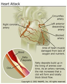
Aortic valve regurgitation develops when the does not function correctly. To understand this condition, it's helpful to know how the aortic valve normally functions. The aortic valve works like a one-way gate, opening so that blood from the left ventricle (the heart's main pump) can be pushed into the , the large artery leaving the heart. From the aorta, oxygen-rich blood flows into the branching arteries and through the body to feed the cells. When the heart rests between beats, the aortic valve closes to keep blood from flowing backward into the heart. See a picture .

In aortic valve regurgitation, the aortic valve does not close properly. With each heartbeat, some of the blood pumped into the aorta leaks back (regurgitates) through the faulty valve into the left ventricle. The body doesn't receive enough blood, so the heart must work harder to make up for it (compensation). See a picture of

Typically, symptoms do not develop for decades because the heart compensates by getting bigger so that it can pump out more blood. But, if it is not corrected, regurgitation usually gets worse over time, and symptoms such as shortness of breath and fatigue develop. At this point, an aortic valve replacement is typically needed to prevent (arrhythmias), and irreversible damage to the heart muscle.

In rare cases, aortic valve regurgitation comes on suddenly and requires immediate medical attention.

Some people have very small amounts of blood that leak back into the left ventricle. This usually doesn't cause any symptoms or problems. This topic focuses on the more serious cases of aortic valve regurgitation where large amounts of blood flow back across the aortic valve into the left ventricle.

What causes aortic valve regurgitation?

Any condition that damages the aortic valve can cause aortic valve regurgitation. Common causes include being born with a defective aortic valve, wear and tear from aging, infection of the lining of the heart and . Enlargement of the aorta, associated with and hardening of the arteries , can also cause aortic valve regurgitation. On rare occasions, to the chest can damage the aortic valve.

Rarer conditions that cause aortic valve regurgitation include a disorder of the body's connective tissues, a type of arthritis, some, and.

The most common causes of sudden (acute) aortic valve regurgitation include:

  • Endocarditis, which is an infection in the heart caused by bacteria.
  • which is the separation of the inner layer of the aorta from the middle layer.
  • Problems with the.

Other conditions that cause acute regurgitation include trauma to the heart valve.

What are the symptoms?

Hypertension: High Blood Pressure and Atherosclerosis

What is atherosclerosis and how is it related to high blood pressure?

One of the most serious health problems related to untreated high blood pressure is atherosclerosis, which contributes to coronary artery disease.

People with high blood pressure are more likely to develop coronary artery disease because high blood pressure puts added force against the artery walls. Over time, this extra pressure can damage the arteries making them more vulnerable to the narrowing and plaque build up associated with atherosclerosis. The narrowed artery limits or blocks the flow of blood to the heart muscle depriving the heart of oxygen. The hardened surface of the artery can also encourage the formation of small blood clots.

What Are the Symptoms of Atherosclerosis?

Atherosclerosis usually has no symptoms until the narrowed coronary arteries severely restrict blood flow to the heart. At this point, you may feel chest pain because not enough blood is reaching the heart. These pains may come with exertion, or can occur when the person is at rest.

What Is a Heart Attack?

A heart attack is permanent damage to the heart muscle caused by loss of blood flow to the heart. A heart attack occurs when a clot or spasm blocks an already narrowed coronary artery. Left without oxygen, the portion of the heart muscle served by the blocked artery is injured.

How Is Atherosclerosis Diagnosed?

Atherosclerosis isn't usually diagnosed until a person complains of chest pain. At this point, the doctor may conduct tests to evaluate your risk for heart disease, including:

  • Electrocardiogram (EKG or ECG): This test measures the electrical activity, rate, and rhythm of your heartbeat via electrodes attached to your arms, legs, and chest. This test may show if you've had a heart attack in the past or if you are currently having a heart attack, strain, or thickening of the heart muscle. It can also convey information about the size of your heart as well. It cannot predict your future risk.
  • Exercise stress test: During this test you exercise on a stationary bicycle or treadmill to increase your heart rate while EKG readings are taken. This test is often performed with imaging of the heart, such as an echocardiogram or nuclear scans, to provide better detection of heart disease.
  • Cardiac catheterization: During this procedure, a catheter -- a small flexible tube -- is inserted into an artery and guided to the coronary arteries, allowing the doctor an opportunity to locate any blockages in the coronary arteries and observe pressure and blood flow in the heart. During a cardiac catheterization, a cardiologist may be able to open up clogged arteries directly from the inside with a procedure called an angioplasty. This may also involve placing a small cylindrical support device in the vessel called a stent.
  • Echocardiogram: This test sends ultrasound waves that provide pictures of the heart's valves and chambers, giving information about chamber size and wall thickness and how blood is flowing within the chambers.
  • Computed tomography (CT) scan: An X-ray and a computer are used to construct a cross-sectional image of the heart.
  • Magnetic resonance arteriography (MRA): This test provides images of blood vessels to show where they may be blocked.

Heart Attacks

More than 1 million Americans have heart attacks each year. A heart attack, or myocardial infarction (MI), is permanent damage to the heart muscle. "Myo" means muscle, "cardial" refers to the heart, and "infarction" means death of tissue due to lack of blood supply.

What Happens During a Heart Attack?

The heart muscle requires a constant supply of oxygen-rich blood to nourish it. The coronary arteries provide the heart with this critical blood supply. If you have those arteries become narrow and blood cannot flow as well as it should. Fatty matter, calcium, proteins, and inflammatory cells build up within the arteries to form plaques of different sizes. The plaque deposits are hard on the outside and soft and mushy on the inside.

When the plaque is hard, outer shell cracks (plaque rupture), platelets (disc-shaped particles in the blood that aid clotting) come to the area, and blood clots form around the plaque. If a blood clot totally blocks the artery, the heart muscle becomes "starved" for oxygen. Within a short time, death of heart muscle cells occurs, causing permanent damage. This is a heart attack.

While it is unusual, a heart attack can also be caused by a spasm of a coronary artery. During a coronary spasm, the coronary arteries restrict or spasm on and off, reducing blood supply to the heart muscle (ischemia). It may occur at rest and can even occur in people without significant

Each coronary artery supplies blood to a region of heart muscle. The amount of damage to the heart muscle depends on the size of the area supplied by the blocked artery and the time between injury and treatment.

Healing of the heart muscle begins soon after a heart attack and takes about eight weeks. Just like a skin wound, the heart's wound heals and a scar will form in the damaged area. But, the new scar tissue does not contract or pump as well as healthy heart muscle tissue. So, the heart's pumping ability is lessened after a heart attack. The amount of lost pumping ability depends on the size and location of the scar.

What Are the Symptoms of a Heart Attack?

Symptoms of a heart attack include:

  • Discomfort, pressure, heaviness, or pain in the chest, arm or below the breastbone
  • Discomfort radiating to the back, jaw, throat or arm
  • Fullness, indigestion, or choking feeling (may feel like heartburn)
  • Sweating, nausea, vomiting, or dizziness
  • Extreme weakness, anxiety, or shortness of breath
  • Rapid or irregular heartbeats

During a heart attack, symptoms last 30 minutes or longer and are not relieved by rest or oral medications (medications taken by mouth).

Some people have a heart attack without having any symptoms (a "silent" myocardial infarction). A silent MI can occur in any person, though it is more common among diabetics

Heart Disease and Heart Bypass Surgery

Heart bypass surgery can be used to treat heart disease when your coronary arteries are blocked. Your doctor may treat the problem by giving the blood a new pathway to the heart.

During coronary artery bypass graft surgery (also called CABG, or "cabbage") a blood vessel is removed or redirected from one area of the body and placed around the area or areas of narrowing to "bypass" the blockages and restore blood flow to the heart muscle. This vessel is called a graft.

These substitute blood vessels can come from your chest, legs, or arms. They're safe to use because there are other pathways that take blood to and from those tissues. The surgeon will decide which graft(s) to use depending on the location of your blockage, the amount of blockage and the size of your coronary arteries.

Heart bypass surgery is the most commonly performed surgery in the U.S.

Which Arteries Are Used for Coronary Grafts?

There are several types of bypass grafts used for heart bypass surgery. The surgeon decides which graft(s) to use, based on the location of the blockage, the amount of blockage, and the size of the patient's coronary arteries.

  • Internal mammary arteries (also called thoracic arteries). These are the most common bypass grafts used, as they have been shown to have the best long-term results. In most cases, these arteries can be kept intact at their origin since they have their own oxygen-rich blood supply. During the procedure, the arteries are sewn to the coronary artery below the site of blockage. This artery is located in the chest and can be accessed through the primary incision for the bypass surgery.
  • Saphenous veins. These veins are removed from your leg, and then sewn from your aorta to the coronary artery below the site of blockage. Minimally invasive saphenous vein removal may be performed and results in less scarring and a faster recovery.
  • Radial artery. There are two arteries in the lower part of the arm, the ulnar and radial arteries. Most people receive adequate blood flow to their arm from the ulnar artery alone and will not have any side effects if the radial artery is removed and used as a graft. Careful preoperative and intraoperative tests determine if the radial artery can be used. If you have certain conditions (such as Raynaud's, carpal tunnel syndrome, or painful fingers in cold air) you may not be a candidate for this type of bypass graft. The radial artery incision is in your forearm, about 2 inches from your elbow and ending about 1 inch from your wrist. If you have this type of bypass, you will probably be prescribed a medication called a for about six months after surgery to help keep the radial artery open. Some people report numbness in the wrist after surgery. However, long-term sensory loss or numbness is uncommon.
  • The gastroepiploic artery to the stomach and the inferior epigastric artery to the abdominal wall are less commonly used for grafting.

It is common for three or four coronary arteries to be bypassed during surgery. A coronary artery bypass can be performed with traditional surgery (see below) or with minimally invasive surgery (see below). Your surgeon will review your diagnostic tests prior to your surgery to see if you are a candidate for minimally invasive bypass surgery.

Angioplasty Versus Bypass Surgery for Coronary Artery Disease

Andrew D. Michaels, MD; Kanu Chatterjee, MB, FRCP

From the Division of Cardiology, Department of Medicine, University of California at San Francisco Medical Center, San Francisco.

Correspondence to: Kanu Chatterjee, MB, FRCP, Professor of Medicine, Lucie Stern Professor of Cardiology, Chatterjee Center for Cardiac Research, University of California, San Francisco, 505 Parnassus Ave, San Francisco, CA 94143. E-mail

What Is Coronary Artery Disease?

Coronary artery disease (CAD) is a disease caused by "hardening" (termed atherosclerosis) of the coronary arteries on the surface of the heart CAD is the leading cause of death for both men and women in the United States.

Didn’t find what you are looking for? Try out Google Search